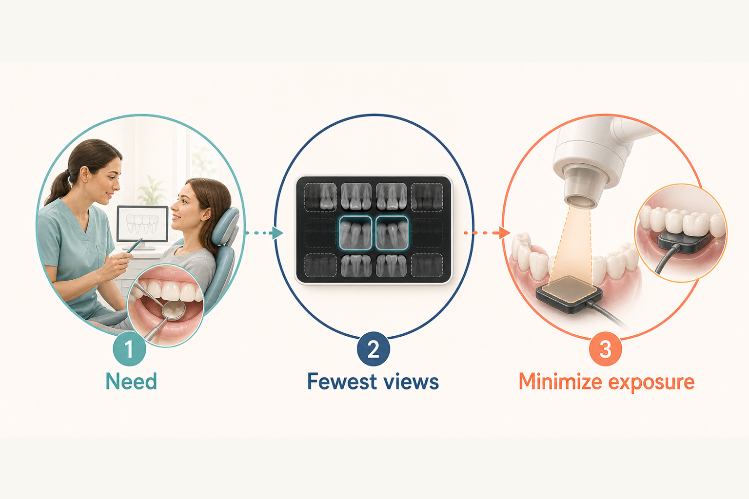
Dental X-rays are generally considered safe when they are needed for diagnosis or treatment planning and the dental team uses current methods to limit exposure. They do involve ionizing radiation and are not completely risk-free, so they should be taken for a clear clinical reason—not automatically at every visit.
The useful question is not whether an X-ray has zero risk. It is whether the expected diagnostic benefit justifies the small exposure and whether the fewest appropriate images can answer the clinical question.
Why dentists use X-rays
A visual examination cannot show every part of a tooth, its roots, or the surrounding bone. Radiographs can provide information about areas that are difficult or impossible to see directly. Depending on the image and the clinical question, they may help a dentist evaluate:
- Decay between teeth or beneath some existing dental work
- Bone levels around teeth
- Tooth roots and the tissues around them
- Developing or unerupted teeth
- Injuries, infections, or other changes that need further assessment
- Planning or follow-up for certain procedures
An X-ray is one piece of the evaluation. Dentists interpret it alongside symptoms, health and dental history, the examination, and previous images.
Common types of dental X-rays
Different images answer different questions.
- Bitewing X-rays show the crowns of upper and lower teeth in the same area and are often used to assess spaces between back teeth and supporting bone.
- Periapical X-rays show an entire tooth, including its root and nearby bone.
- Panoramic X-rays create a broad view of the jaws, teeth, and surrounding structures.
- Occlusal X-rays show a larger area of one arch and may help assess tooth development or location.
- Cone-beam computed tomography (CBCT) creates three-dimensional information for selected situations. It generally exposes a patient to more radiation than routine two-dimensional dental images, so it should be used only when that added information is expected to matter.
Not every patient needs every type.
What “low exposure” means
Dental radiographs use ionizing radiation. Any radiation exposure should be taken seriously, but risk depends on the dose and the circumstances. Modern dental imaging is designed to obtain diagnostically useful information with low exposure.
It is easy to find dramatic comparisons online, but exact doses vary with the image type, machine, settings, receptor, beam size, and technique. That is why a single comparison cannot describe every dental X-ray.
Digital receptors can reduce exposure compared with slower film systems when equipment and settings are used appropriately. Digital technology does not make an unnecessary image necessary, however; clinical justification remains the first safety step.
How exposure is minimized
Radiation safety begins with deciding whether an image is likely to help. Current ADA guidance emphasizes that radiographs should be ordered only when clinically indicated and that exposure should be kept as low as diagnostically acceptable.
Practical safeguards can include:
- Reviewing prior images so useful records are not repeated without reason
- Choosing the image type and number needed for the question
- Using appropriate digital receptors or fast film
- Limiting the X-ray beam to the area being imaged
- Selecting exposure settings appropriate for the patient and task
- Positioning equipment carefully to reduce retakes
- Maintaining and testing imaging equipment
You may hear the term ALARA, meaning “as low as reasonably achievable.” Newer guidance also uses ALADA, “as low as diagnostically acceptable,” to stress that an image still has to be good enough to answer the clinical question.
Why X-rays are not taken at every visit
There is no one schedule that fits everyone. The need and timing can depend on age, symptoms, cavity risk, gum health, dental history, current findings, past treatment, tooth development, and the availability and quality of previous images.
A person with new symptoms or a higher risk of disease may need images sooner than someone with no symptoms, a low risk, and recent usable records. Routine screening images should not replace a clinical history and examination.
If a new image is recommended, reasonable questions include:
- What are you looking for?
- How could the result change what happens next?
- Are recent images from another office available?
- Which type and how many images are needed?
Pregnancy considerations
Tell the dental team if you are pregnant or might be pregnant. This gives them the context to confirm that an image is needed and select appropriate techniques.
Professional guidance supports taking clinically necessary dental radiographs during pregnancy rather than delaying needed diagnosis or care solely because of pregnancy. The decision should still be individualized. A patient and dental team can discuss why the image is recommended, whether it can reasonably wait, and what exposure-limiting measures will be used.
Recommendations about lead aprons and thyroid collars have changed as equipment and evidence have evolved, and local regulations may differ. The ADA’s 2024 expert guidance does not recommend routine abdominal or thyroid shielding during dental imaging. Shielding can interfere with the image or equipment and sometimes lead to a retake; selecting only necessary images, limiting the beam, and using appropriate settings are more important exposure-reduction measures. An office may follow different requirements based on state rules or specific circumstances.
Are digital X-rays safer?
Digital imaging often allows a useful image with less radiation than traditional film, especially compared with slower film. It also makes images easier to store, enlarge, and share. Safety still depends on appropriate selection, equipment, settings, positioning, and quality control. “Digital” alone is not a guarantee that every exposure is optimized.
The takeaway
Dental X-rays have a small radiation risk, but they can reveal information that a direct examination cannot. They are considered appropriate when the expected benefit outweighs that risk and the dental team obtains only the images needed with exposure-minimizing methods.
They should not be an automatic part of every appointment. At a dental checkup, the recommendation should reflect your history, examination, risk, prior records, and the question the image is meant to answer.
Sources
- X-Rays/Radiographs — American Dental Association
- Optimizing radiation safety in dentistry — American Dental Association
- ADA Releases Updated Recommendations to Enhance Radiography Safety in Dentistry — American Dental Association
- The Selection of Patients for Dental Radiographic Examinations — U.S. Food and Drug Administration
- Medical X-ray Imaging — U.S. Food and Drug Administration