The simple map
A tooth has two main regions: the crown, which is normally visible above the gumline, and the root, which sits below the gumline and is held in the jaw. Within those regions are several tissues, including enamel, dentin, cementum, and pulp.
Dentists use these anatomy words because location gives context to a finding. You do not need to memorize every term. A simple map can help you follow the conversation and ask clearer questions.
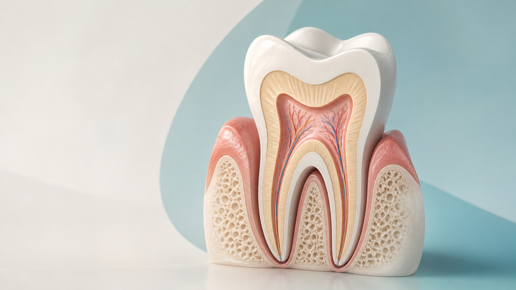
The illustration is a simplified teaching picture. Real teeth vary in shape and number of roots, and microscopic structures are not shown to scale.
Crown, root, neck, and gumline
The crown is the part of the tooth you usually see. The root is the part below the crown that is held in its socket. The neck is where the crown and root meet. The gumline describes the edge where the gum meets the tooth.
These words describe location, not a diagnosis. For example, a dentist may talk about a change on a chewing surface, a crack near an old filling, or an area close to the gumline.
Enamel covers the crown
Enamel is the hard outer covering of the crown. It protects the dentin beneath it. Acids produced by mouth bacteria can cause enamel to lose minerals over time.
Early tooth decay usually causes no symptoms. A dentist may notice a change during an examination or on an x-ray before you feel anything. That does not mean every mark or sensation is decay; appearance and symptoms alone cannot confirm the cause.
Dentin is underneath enamel
Dentin lies under the enamel in the crown and under cementum along the root. It contains microscopic tubules. When dentin loses its protective covering, heat, cold, or certain foods can stimulate tissues inside the tooth and contribute to sensitivity.
Sensitivity still has more than one possible cause. The pattern can offer useful context, but a symptom cannot show you which layer is involved. For a closer look at that distinction, see why teeth may hurt with cold food or drinks.
Pulp is the living tissue inside
Pulp is the soft tissue in the center of the tooth. It contains nerves, blood vessels, and connective tissue. The pulp chamber is in the crown, and the pulp space continues down through the root canals.
When a dentist says that a finding is “close to the nerve,” they are usually describing its relationship to the pulp. Whether the pulp is healthy or inflamed requires a clinical assessment; symptoms alone cannot establish that. If this term comes up during a treatment discussion, our root canal basics guide explains the broader context without predicting what treatment you need.
Cementum and the periodontal ligament help hold the root
The root is not covered by enamel. Cementum is the hard tissue that covers the root. The periodontal ligament is a network of connective-tissue fibers between the root and its socket. Together with the surrounding bone, these structures help support the tooth.
These tissues are easy to leave out of a simple picture, but the distinction matters: crown and root surfaces have different coverings.
Bone and gums support the tooth
Teeth do not sit in the mouth by themselves. Gingiva, the clinical word for gums, covers and protects the tissues around the teeth. Alveolar bone, the tooth-supporting part of the jawbone, surrounds the roots and forms their sockets.
A dentist may evaluate a tooth together with the gums, supporting tissues, bite, x-rays, and dental history. Root exposure, gum inflammation, and bone changes are different findings even though they occur in the same small area.
How anatomy words add context to a finding
| Term you may hear | What it locates | What it does not tell you by itself |
|---|---|---|
| “In the enamel” | The hard outer covering of the crown | Whether a visible spot is active decay or what care is appropriate |
| “Into the dentin” | The layer beneath enamel or cementum | Why you have a particular symptom |
| “Near the pulp” | The finding is close to the tooth’s living center | Whether the pulp is inflamed or which treatment you need |
| “On the root surface” | The area below the crown, covered by cementum rather than enamel | Why the root is exposed or sensitive |
| “Around the supporting bone” | The tissues that hold the tooth in its socket | The cause or significance of a change |
This vocabulary can also make questions more specific: “Which part of the tooth are you seeing?” “How was the depth estimated?” and “What other findings affect the recommendation?”
The main idea
Tooth anatomy gives you a shared vocabulary for dental visits. Crown, root, enamel, dentin, cementum, pulp, periodontal ligament, gums, and bone describe different places and tissues. They do not diagnose a problem or determine treatment by themselves.
If you are trying to understand pain or a possible cavity, continue with what tooth pain can mean or what a cavity may look and feel like. A dentist can combine what you notice with an examination and, when appropriate, dental imaging.
Sources
- Tooth — MouthHealthy, American Dental Association
- Tooth Decay — National Institute of Dental and Craniofacial Research